animal cell microscope labeled
As you can see in the above labeled plant cell diagram under light microscope there are 13. Learn vocabulary terms and more with flashcards games and other study tools.
Here S How Plant And Animal Cells Are Different Howstuffworks
What are plant and animal cells called.

. One vital part of an animal cell is the nucleus. As you can see in the above labeled plant cell diagram under light microscope there are 13 parts namely Cell membrane. Animal Cell - Science Quiz.
Find and download Labeled Animal Cell Electron Microscope image wallpaper and background for your Iphone Android or PC Desktop. Generalized Structure of a Plant Cell Diagram. Start studying BIOLOGY MICROSCOPE SLIDES ANIMALPLANTBACTERIA CELLS LABELED AND MORE EXAM.
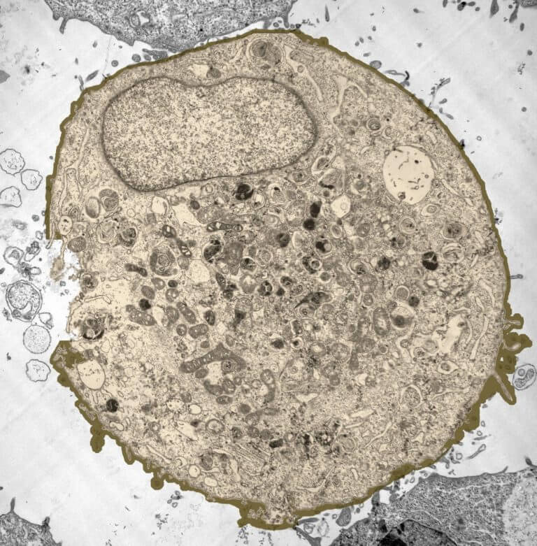
Cheek cell drawing any power but preferably high Drawings Conclusions and Questions. Labeled animal cell under electron microscope. The microscope parts work together in hospitals and in forensic labs for scientists and students bacteriologists and biologists so that they may view bacteria plant and animal.
Realtec have about 24 image published on this page. Typical Animal Cell Pinocytotic vesicle Lysosome Golgi vesicles Golgi vesicles rough ER endoplasmic reticulum Smooth ER no ribosomes Cell plasma membrane. You get the best of both worlds.
Most cells both animal and plant range in size between 1 and 100 micrometers and are thus visible only with the aid of a microscope. Animal cells are packed with amazingly specialized structures. The animal cell is more fluid or elastic or.
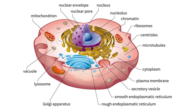
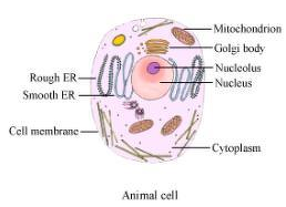
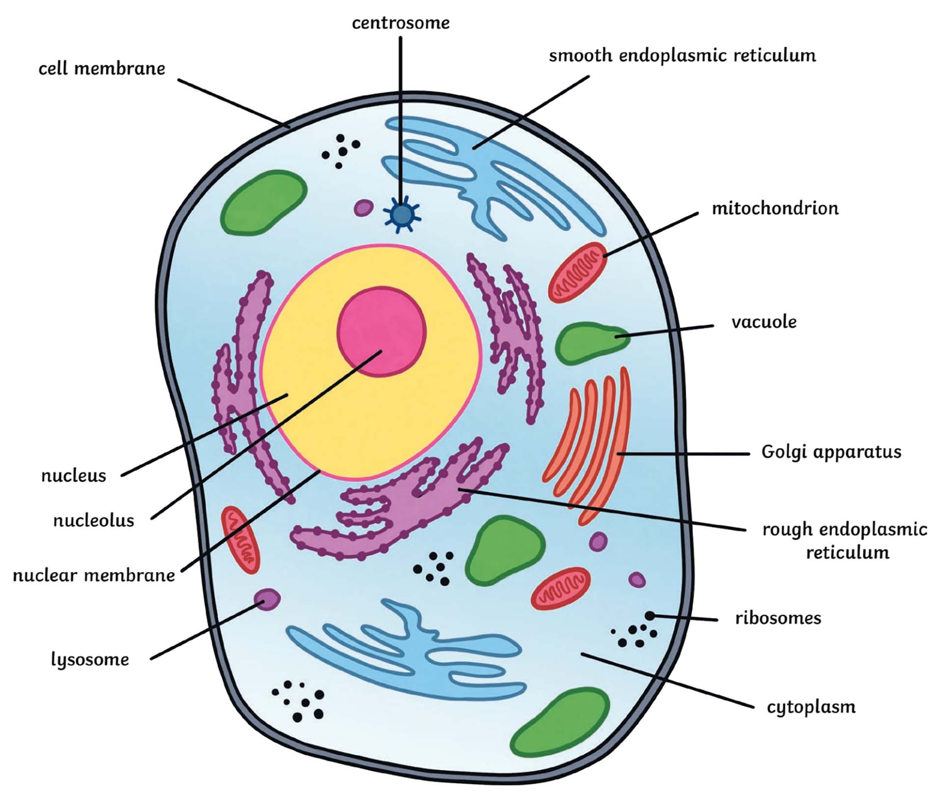
There are 13 main parts of an animal cell. General microscope handling instructions hold with one hand 13 fresh labeled animal cell under electron microscope. Animal cells are eukaryotic cells that contain a membrane-bound nucleus.
We hope this detailed article on Plant and. Diagram Of Animal Cell. Animal Cell Diagram Under Microscope Labeled.
Your microscope has four objectives of varying magnifications 4x 10x 40x and 100x mounted on a revolving. Onion Cell drawing high power 2. Draw a large diagram of an animal cell as seen through an.
Animal cells have a basic structure. Its the cells brain employing chromosomes to. For viewing under the light microscope can label plant and animal cell structures and describe their functions to be able.
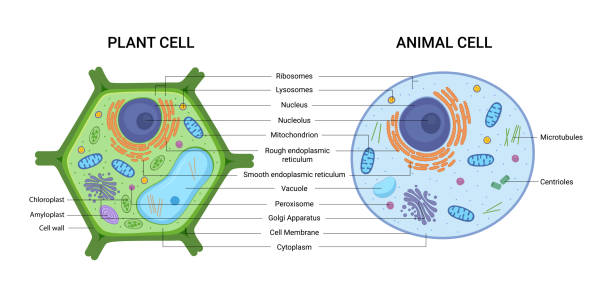
They are different from plant cells in that they do contain cell walls and chloroplast. Cell membrane nucleus nucleolus nuclear membrane cytoplasm endoplasmic reticulum Golgi apparatus. The lack of a rigid cell wall allowed.
Explore topics on usage care terminology and. Neuron under microscope labelled diagram. Plant and animal cells are called Eukaryotic because the true nucleus is present.
Animal Cell Microscope Labeled.
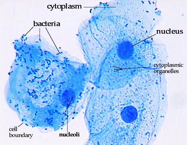
What Organelles Would Be Visible In A Cheek Cell Why Quora
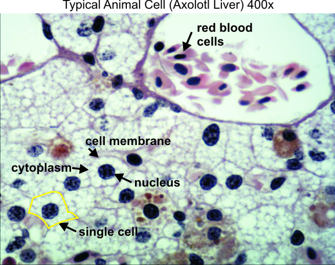
Typical Animal Cell 400x Dissection Connection

1 2 Difference Between Plant And Animal Cells Cells As The Basic Units Of Life Siyavula
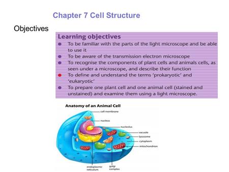
Animal Plant Cells Lo To Be Able To Prepare Slides For Viewing Under The Light Microscope Can Label Plant And Animal Cell Structures And Describe Their Ppt Download
Amazing 27 Things Under The Microscope With Diagrams
Q14 Draw A Large Diagram Of An Animal Cell As Seen Through An Electron Microscope Label The Parts That Science Tissues 11500353 Meritnation Com
What Are Cells Animal And Plant Cells Ks3 Biology Bbc Bitesize Bbc Bitesize

Q14 Draw A Large Diagram Of An Animal Cell As Seen Through An Electron Microscope Label The Parts Brainly In
Amazing 27 Things Under The Microscope With Diagrams

Lab Manual Exercise 1a

Acg055 General Animal Cell Valley Microscope
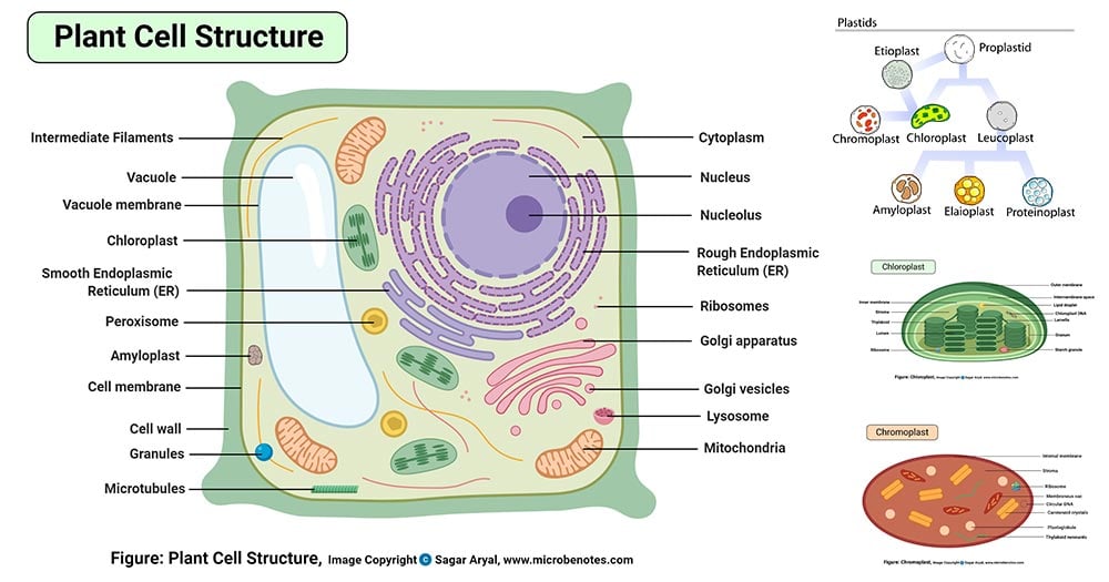
Plant Cell Definition Structure Parts Functions Labeled Diagram
What Is An Animal Cell Definition And Functions Twinkl
Amazing 27 Things Under The Microscope With Diagrams

Gce Cie Biology Animal And Plant Cell Structures And
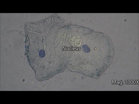
Cheek Cells Under The Microscope Youtube

Cellular Biology And Microscopy Ppt Download Animal Cell Structure Cell Organelles Organelles
7 202 Plant Cell Illustrations Clip Art Istock

Biology Cells And Microscope Diagram Quizlet